
Fig. 3
- ID
- ZDB-FIG-210426-14
- Publication
- Duszyc et al., 2021 - Mechanotransduction activates RhoA in the neighbors of apoptotic epithelial cells to engage apical extrusion
- Other Figures
- All Figure Page
- Back to All Figure Page
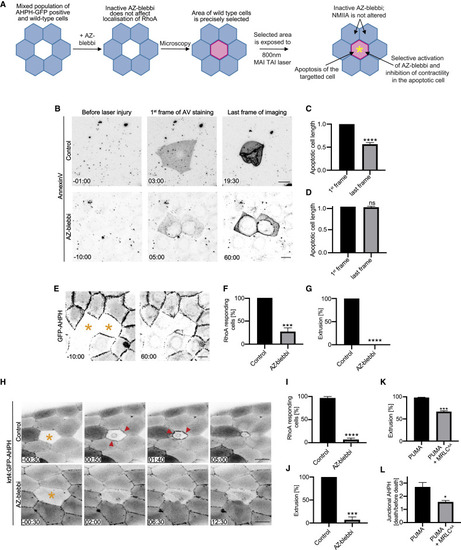
Contractility in the apoptotic cell is necessary for it to activate RhoA in its neighbors (A) Schematic diagram of the experimental design for simultaneous activation of azido-blebbistatin (AZ-blebbi) and induction of apoptosis in targeted cells. (B–D) Azido-blebbistatin allows contractility to be inhibited in apoptotic cells. (B) Morphological changes when apoptosis was sporadically induced by laser microirradiation in confluent control and azido-blebbistatin-treated monolayers. To quantitate contractility, we measured the longest axis of the apoptotic cells for controls (C) and azido-blebbistatin (20 μM)-treated cultures (D) from when annexin-V labeling was first evident (1st frame) till either extrusion was completed (for controls, last frame) or 60 min after injury (AZ-blebbi, last frame). (E–G) Activation of azido-blebbistatin in apoptotic cells prevents activation of RhoA in neighbors and inhibits extrusion. (E) Representative montage of GFP-AHPH in neighbors of apoptotic cells where azido-blebbistatin had been simultaneously activated (asterisks). (F and G) Activation of (F) RhoA in neighbors, measured as the percentage of cells that showed a preferential increase in GFP-AHPH at the apoptotic:neighbor interface and (G) apoptotic extrusion. See also Figure S3F. (H–J) Activation of azido-blebbistatin in apoptotic cells in zebrafish periderm prevents activation of RhoA in neighbors and inhibits extrusion. (H) Representative montage of GFP-AHPH in neighbors of apoptotic cells (asterisk) in control (top panels) and where azido-blebbistatin had been simultaneously activated (asterisk, bottom panels). (I and J) Activation of (I) RhoA in neighbors, measured as the percentage of cells that showed a preferential increase in GFP-AHPH at the apoptotic:neighbor interface and (J) apoptotic extrusion. (K and L) Inhibition of apoptotic contractility prevents extrusion (K) and activation of RhoA in neighbors (L) when apoptosis was induced in Caco2 cells by PUMA. Apoptotic cell contractility was inhibited by co-expression of inhibitory MRLCAA in the PUMA cells. Junctional AHPH was quantified as the fold change of fluorescence intensity at the frame when apoptosis was confirmed by Draq7 staining relative to the intensity of AHPH before apoptosis. See also Figures S3I and S3J. Scale bars represent 15 μm. All data are means ± SEM; ns, not significant; ∗p < 0.05; ∗∗∗p < 0.001; ∗∗∗∗p < 0.0001; calculated from n = 3 independent experiments analyzed with Student’s t test. Time is mm:ss. XY panels present maximum projection of all acquired z stacks. |