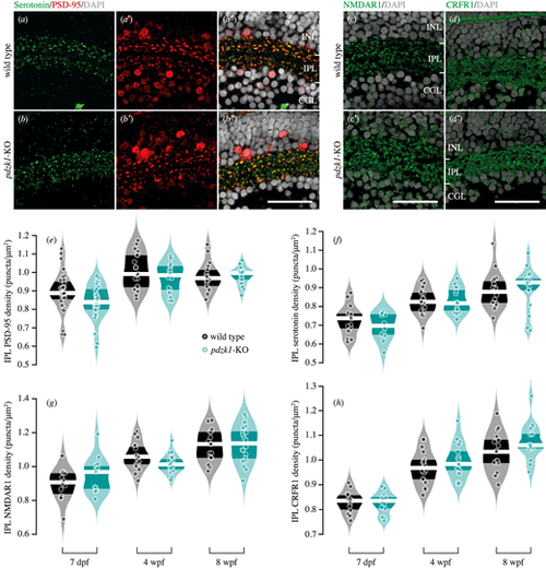
Immunostaining and density quantification of PDZK1-interacting molecules in wild-type and pdzk1-KO retinas. Micrographs of retinal sections from 7-dpf wild-type and pdzk1-KO larvae were co-labeled with (a–b″) serotonin and PSD-95 in green and red, respectively. (c, c′) NMDAR1 and (d, d′) CRFR1 were labeled in green for 7-dpf retinas. For all images, nuclei were stained with DAPI, shown in gray. Scale bars: 25 µm. Violin plots show densities of (e) PSD-95, (f) serotonin, (g) NMDAR1, and (h) CRFR1 in the IPL at 7 dpf, 4 wpf, and 8 wpf. Points are data from individual retinas. White lines represent medians, and dark bands indicate interquartile ranges. Statistical comparisons were performed using two-way ANOVA with Bonferroni correction and Bayesian ANOVA. There were between 12 and 31 retinas per group (see Supplementary information). BFinclusion < 0.33 was considered to be evidence for the null hypothesis (i.e., the data are at least three times as likely under the null hypothesis than the alternative).
|
