
Fig. 1
- ID
- ZDB-FIG-210422-43
- Publication
- Xie et al., 2021 - Altered Visual Function in a Larval Zebrafish Knockout of Neurodevelopmental Risk Gene pdzk1
- Other Figures
- All Figure Page
- Back to All Figure Page
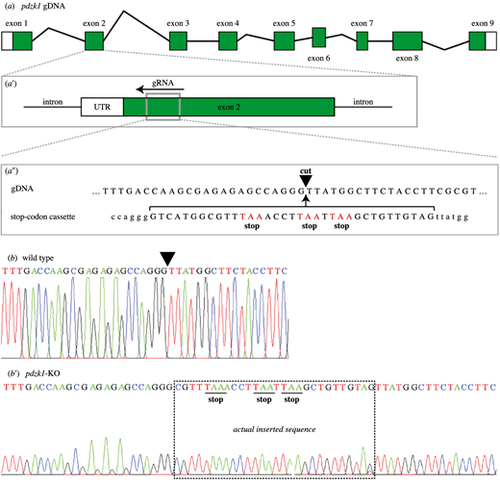
CRISPR editing of pdzk1. (a) There are nine exons in pdzk1 genomic DNA. (a′) The guide RNA (gRNA) targets a site in exon 2 (gray box) and recruits the Cas9 enzyme to recognize and cut the DNA. (a″) The target cut site (black inverted triangle) has homologous arms matching the stop codon template that is co-injected with Cas9 and gRNA during the one-cell stage of zebrafish development. In this way, homology-directed repair inserts the stop codon cassette at exon 2 of pdzk1. Stop codons of the injected cassette are highlighted in red. (b) Sequence results for wild-type fish. The inverted triangle indicates the targeted cut site. (b′) Sequence results for homozygous pdzk1-knockout fish. The box shows the sequence inserted by CRISPR gene editing including the three stop codons (underlined). Sequences on either side were identical to the wild-type fish. |