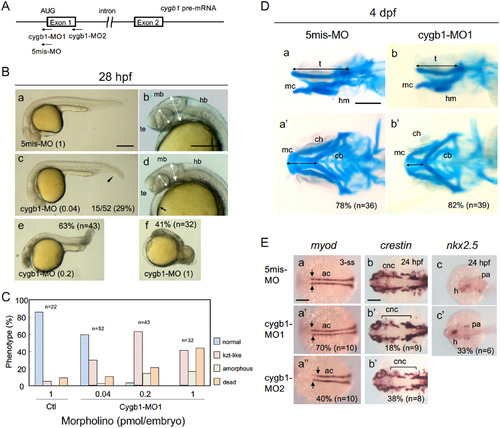
Knockdown of cygb1 phenocopied the kzt mutation. (A) Morpholino oligos were designed against the translational start codon (cygb1-MO1) and the donor site at the exon 1-intron 1 junction (cygb1-MO2). As a negative control, cygb1-MO1 with five mismatches was used in parallel (5mis-MO). (B) Embryos injected with 5mis-MO at 1 pmol/embryo (a, b) and cygb1-MO1 at 0.04 pmol/embryo (c, d), 0.2 pmol/embryo (e), and 1 pmol/embryo (f) were observed at 28 hpf. cygb1-MO1 at 0.04 pmol/embryo caused head flattening (two-headed arrows), small eyes (two-headed broken arrows), trunk-tail bending (arrowhead), and heart edema (arrow), thus phenocopying the kzt mutation. A higher concentration (1 pmol/embryo) resulted in phenotypes much more severe than those of kzt embryos, whereas 5mis-MO1 resulted in few anomalies. (C) Dose-dependent effects of cygb-MO1 on development. Embryos with normal morphology, kzt-phenotypes, amorphous embryos, and death were scored at different doses of cygb1-MO1. (D) Development of the cranial cartilage in embryos injected with morpholino oligos. Cranial cartilage was stained with Alcian blue in embryos injected with 5mis-MO (a, a’) or cygb1-MO1 (b, b’). (E) Knockdown of cygb1 resulted in molecular effects in embryos similar to those of the kzt mutation. The expression of myod in adaxial cells and crestin in NCCs was examined in embryos injected with 5mis-MO (a–c), cygb1-MO1 (a’–c’), or cygb1-MO2 (a’’, b’’). The pattern of myod expression was particularly affected in the anterior adaxial cells (arrows), and crestin expression was particularly decreased in pharyngeal arch-derived cartilage (brackets). (B, Da, Db) Lateral views with anterior to the left and dorsal to the top. (Da’, Db’, E) Dorsal views with anterior to the left. Percentages of abnormal embryos and the numbers of embryos examined are shown at the bottom right or at the top right. ac, adaxial cell; ch, ceratohyal; cb, ceratobranchial; cnc, cranial neural crest cell; h, heart; hb, hindbrain; hm, hyomandibular; mb, midbrain; mc, Meckel’s cartilage; pa, pharyngeal arch; t, trabecula, te, telencephalon. Scale bars, 200 μm (B, E) or 100 μm (D).
|
