
Fig. 2
- ID
- ZDB-FIG-210414-72
- Publication
- Takahashi et al., 2020 - A globin-family protein, cytoglobin 1, is involved in the development of neural crest-derived tissues and organs in zebrafish
- Other Figures
- All Figure Page
- Back to All Figure Page
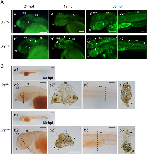
Increased cell death in the central nervous system of kzt embryos. (A) Acridine orange (AO) staining shows abundant cell death in kzt embryos. Offspring obtained by mating kzt heterozygotes were stained at 24 hpf, 48 hpf, and 60 hpf with AO, which stains apoptotic cells as bright spots. Lateral views of the heads (a, b, c1, a’, b’, c1’) and trunk-tail regions (c2, c2’) of kzt+/and kzt–/– embryos are shown, anterior to the left and dorsal to the top. Cell death was detected in the midbrain (mb), hindbrain (hb), optic vesicles (eye, e), telencephalon (te), and spinal cord (sc) of kzt homozygotes (arrows). During the course of development, AO staining became progressively more striking in kzt embryos, whereas it began to be weakly detected in wild-type siblings only after 60 hpf. (B) Terminal deoxynucleotidyl transferase dUTP nick end labeling (TUNEL) confirmed the increased apoptotic cells in kzt embryos. kzt–/– or sibling embryos (kzt+/) were stained at 60 hpf using the TUNEL method. Lateral views of whole embryos (a1, b1), heads (a2, b2), and the trunk-tail region (a3, b3) are shown, with anterior to the left. (a2’, a3’, b2’, b3’) Cross-sections of stained embryos were from the positions marked in lateral views with dotted lines (a2, a3, b2, b3, respectively). As with AO staining, apoptotic cells were intensely detected only in kzt homozygotes (arrowheads in cross-sections). cf, caudal fin; e, optic vesicle; hb, hindbrain, mb, midbrain; sc, spinal cord; te, telencephalon; ∗, notochord. Scale bars, 200 μm. |
| Fish: | |
|---|---|
| Observed In: | |
| Stage Range: | Prim-5 to Pec-fin |
Reprinted from Developmental Biology, 472, Takahashi, K., Ito, Y., Yoshimura, M., Nikaido, M., Yuikawa, T., Kawamura, A., Tsuda, S., Kage, D., Yamasu, K., A globin-family protein, cytoglobin 1, is involved in the development of neural crest-derived tissues and organs in zebrafish, 1-17, Copyright (2020) with permission from Elsevier. Full text @ Dev. Biol.