
Fig. 3
- ID
- ZDB-FIG-210414-73
- Publication
- Takahashi et al., 2020 - A globin-family protein, cytoglobin 1, is involved in the development of neural crest-derived tissues and organs in zebrafish
- Other Figures
- All Figure Page
- Back to All Figure Page
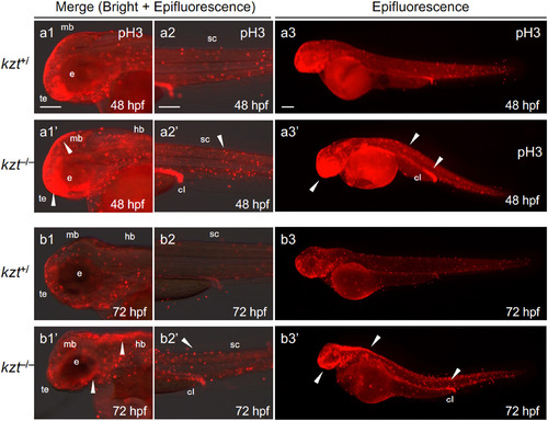
Cell proliferation is increased in kzt embryos. kzt homozygotes (kzt–/–) or sibling embryos (kzt+/) were immunostained for phosphorylated histone H3 (pH3) at 48 hpf or 72 hpf. Epifluorescence images or those merged with bright-field images are shown. Images are lateral views of the heads (a1/a1’, b1/b1’), trunk-tail regions (a2/a2’, b2/b2’), and whole embryos (a3/a3’, b3/b3’), with anterior to the left and dorsal to the top. Higher numbers of proliferative cells (stained red) were found throughout kzt homozygote embryos (arrowheads), including the brain at both 48 hpf and 72 hpf. cl, cloaca; e, optic vesicle; hb, hindbrain, mb, midbrain; sc, spinal cord; te, telencephalon. Scale bars, 200 μm. |
| Fish: | |
|---|---|
| Observed In: | |
| Stage Range: | Long-pec to Protruding-mouth |
Reprinted from Developmental Biology, 472, Takahashi, K., Ito, Y., Yoshimura, M., Nikaido, M., Yuikawa, T., Kawamura, A., Tsuda, S., Kage, D., Yamasu, K., A globin-family protein, cytoglobin 1, is involved in the development of neural crest-derived tissues and organs in zebrafish, 1-17, Copyright (2020) with permission from Elsevier. Full text @ Dev. Biol.