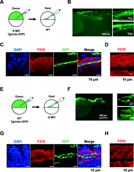
IK functions in a non-cell-autonomous manner in muscle precursors in zebrafish. a Schematic diagram of the cell transplantation. ik morpholino (MO) is injected into Tg (mito: GFP) donor embryo at the one-cell stage. At 4 hpf, the green fluorescent ik MO donor cells are transplanted into WT host embryo. b GFP expression in a chimeric WT host embryo at 36 hpf (left panel). High magnification image of the GFP-labeled cells located in the skeletal muscle (right panel). c Confocal images of chimeric WT host embryo at 36 hpf stained with anti-F310 antibody for fast-twitch muscle fibers and anti-GFP antibody for green fluorescent cells transplanted from ik MO donor embryos. d Confocal images of fast-twitch muscle fibers of WT embryos at 36 hpf stained with anti-F310 antibody. e Schematic diagram of cell transplantation. ik MO is injected into WT host embryo at the one-cell stage. At 4 hpf, the green fluorescent WT Tg (mito: GFP) donor cells are transplanted into ik MO host embryo. f GFP expression in a chimeric ik MO host embryo at 36 hpf (left panel). High magnification image of the GFP-labeled cells located in the skeletal muscle (right panel). g Confocal images of chimeric ik MO host embryo at 36 hpf stained with anti-F310 antibody for fast-twitch muscle fibers and anti-GFP antibody for green fluorescent cells transplanted from WT donor embryo. h Confocal images of fast-twitch muscle fibers of ik MO embryo at 36 hpf stained with anti-F310 antibody
|
