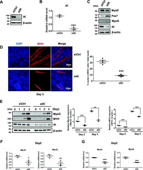
IK-depleted myoblasts have a reduced ability to form normal myotubes. a Immunoblot analysis of IK from C2C12 myoblasts transfected with siIK for 48 h. b Relative mRNA levels of IK from C2C12 myoblast transfected with siIK for 48 h using qRT-PCR analysis. 18S rRNA was used as a normalization control. ***p < 0.001. c Immunoblot analysis of Pax7, MyoD, and MyoG from C2C12 myoblasts transfected with siIK for 48 h. d Immunocytochemistry image stained with anti-MYH antibody from IK-depleted C2C12 cells at day 3 of differentiation. Scale bar = 20 μm (left panel). For fusion index for myotubes, the ratio of the number of nuclei in MYH-positive myotubes per the total nuclei in one field was quantified from four random microscopic fields; (right panel) ***p < 0.001. e Immunoblot analysis of MyoG and MYH from C2C12 cells transfected for 18 h and harvested at days 0, 1, 2, and 3 after differentiation (left panel). The band intensity normalized to β-actin was graphed by ImageJ software (right panel). **p < 0.01, ***p < 0.001. f, g Relative mRNA levels of MyoD and MyoG from C2C12 myoblast f at day 2 and g day 3 after differentiation using qRT-PCR analysis. 18S rRNA was used as a normalization control. *p < 0.05, **p < 0.01, ***p < 0.001
|
