
Fig. 4
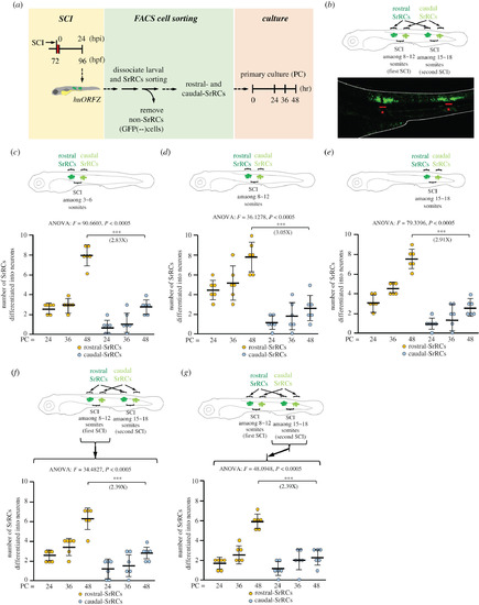
Assessment of neuronal differentiation capability showed that rostral-SrRCs achieved a higher fold increase than caudal-SrRCs during neuronal regeneration following SCI. (a) Schematic diagram illustrating the experimental design. (b) SCI was performed twice simultaneously at 8–12 and 15–18 somites. As demonstrated in the lower panel, a cluster of GFP-expressing SrRCs appeared at both sides of each SCI site, as observed under confocal microscopy. Stars combined with underscore indicate SCI sites. (c–e) Illustration of experimental design by performing single SCI at (c) 3–6, (d) 8–12 and (e) 15–18 somites. The numbers of collected rostral- and caudal-SrRCs able to differentiate into neurons after culturing were calculated separately, and the cell numbers after 48 h culture were compared by the increase in fold. (f,g) Illustration of experimental design by performing SCI simultaneously at 8–12 (the first SCI) and 15–18 somites (the second SCI). The numbers of collected rostral- and caudal-SrRCs from (f) the first 8–12 somite-SCI and (g) the second 15–18 somite-SCI sites able to differentiate into neurons after culturing were calculated separately, and cell numbers after 48 h culture were compared by the increase in fold. Two-way ANOVA with Bonferroni multiple comparisons test, ***p < 0.005; t-test: ***p < 0.001; each F value was also indicated. Error bars indicate s.e.m. |