
Fig. 2
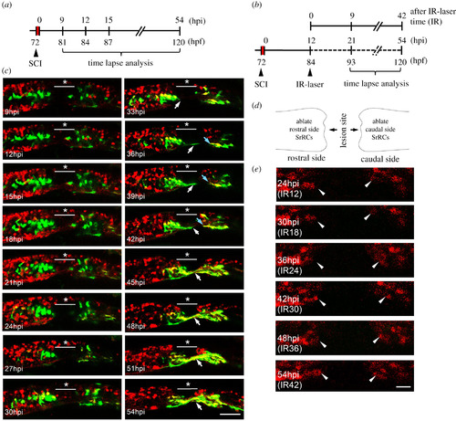
Using an in vivo system to confirm that SCI-induced SrRCs played a role in neuronal regeneration. (a) Schematic diagram of the timetable designed for observing the neuronal regeneration of SCI-larva. hpi: hours-post-injury; hpf: hours-post-fertilization. (b) Schematic diagram of the time table designed for observing the neuronal regeneration of SCI-larvae after infrared (IR)-LEGO laser (IR) treatment which was used to ablate their GFP-expressing cells. (c) Double-transgenic embryos derived from line Tg(huc-DsRed), crossed with line huORFZ, were used to dynamically trace neural differentiation and regeneration following SCI by confocal time-lapse photography. The underscored stars indicate the location where SCI was performed, while white arrows point to cells differentiated into neurons and their extended axons at the series time point indicated. (d) Diagram illustrated that SrRCs located at both sides (rostral and caudal) of SCI site were ablated. (e) Tracing neuronal differentiation and regeneration at series time point, as indicated, after both sides of SrRCs of SCI-larvae were ablated simultaneously. Photos were taken from lateral view in which dorsal side was upward, while rostral side was at left. Arrowheads indicate outgrowing neurites. Scale bar at right bottom was presented as 10 µm. |