
Fig. 1
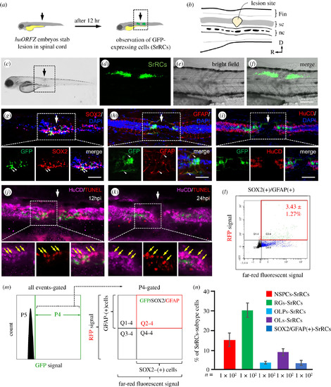
A specific subtype-cell population found in spinal cord of zebrafish larvae following SCI was identified. (a) Schematic diagram of the experiments following SCI. Arrow indicates the spinal cord (sc) injury site. (b) The enlarged image of SCI site shown in the box in (a). nc: notochord. (c) Zebrafish embryos from transgenic line huORFZ at 72 hours-post-fertilization (hpf) were treated with SCI, as indicated by the arrow. Enlarged image of the box area indicated in (c) was observed under microscopy: (d) fluorescence, (e) bright-field and (f) merged images, in which GFP-expressing cells (SrRCs) were apparent on both sides of the SCI site at 12 h post-injury. Scale bar indicates 50 µm. (g–i) Immunostaining identified cell types among the subtype cell population of SrRCs. White arrow indicates the location of SCI site. Red fluorescence was specifically labelled as (g) SOX2, (h) GFAP and (i) HuCD. Yellow fluorescence signal indicated that GFP-expressing SrRCs overlapped target protein labelled by red fluorescence signal, as indicated by yellow arrows. Scale bar indicates 15 µm. (j,k) TUNEL assay identified apoptotic cells by red fluorescence signal. Neurons were identified by marker HuCD labelled with far-red fluorescence signal. The apoptotic neurons appeared in pink colour. (l,m) After FACS, the isolated SrRCs were immunostained with antibodies against SOX2 and GFAP (P5: background; P4: cells expressing GFP signal; Q2–4: GFP(+) cells co-expressing SOX2-infrared light signal and GFAP-RFP signal). (n) Calculating the ratio of each cell type among GFP-expressing cell populations isolated by FACS. Error bars indicate s.e.m. |