
FIGURE
Fig. 4
- ID
- ZDB-FIG-210203-12
- Publication
- Huang et al., 2020 - GFP expression pattern in pituitary and gonads under the control of nuclear progesterone receptor promoter in transgenic zebrafish
- Other Figures
- All Figure Page
- Back to All Figure Page
Fig. 4
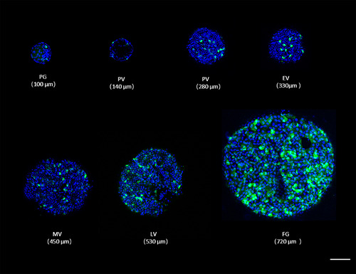
Cellular localization of green fluorescent protein (GFP) under the control of pgr promoter in ovarian follicles during folliculogenesis in Tg(pgr:eGFP). The number of GFP expressing cells increases during folliculogenesis. The diameter of the ovarian follicle was indicated within brackets. The blue fluorescence represented the nucleus which is stained by Hoechst33342. Scale bars = 100 μm. eGFP, enhanced green fluorescent protein; EV, early‐vitellogenic; FG, full‐grown stage; LV, late‐vitellogenic; MV, mid‐vitellogenic; PG, primary growth; Pgr, progesterone receptor; PV, pre‐vitellogenic |
Expression Data
Expression Detail
Antibody Labeling
Phenotype Data
Phenotype Detail
Acknowledgments
This image is the copyrighted work of the attributed author or publisher, and
ZFIN has permission only to display this image to its users.
Additional permissions should be obtained from the applicable author or publisher of the image.
Full text @ Dev. Dyn.