|
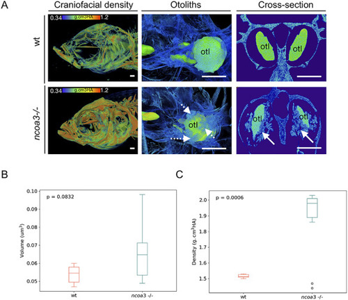
Abnormal mineralisation of amorphous material within the adult inner ears and higher BMD in ncoa3−/−. A) 3D renders from μCT images of wt and ncoa3−/− of same age (1 year old). The head was color-coded to show bone mineral density (g.cm3HA; min = 0.338; max = 1.124). Note that craniofacial bones in ncoa3 mutants have higher density compared to wt. Otoliths (otl = arrows) were zoomed in. Abnormal mineralisation (dashed arrows) is observed attached to the otoliths. A cross section picture was taken to show the mineralised amorphous material (arrows) juxtaposed to the otoliths. B) Volume of otoliths. C) Bone mineral density of central region of otoliths. Non-parametric, two-tailed, independent Student’s t-Test was used as statistical analysis (p < 0.05). Scale bars = 500 μm.
|