IMAGE

Figure 4

ID
ZDB-IMAGE-210130-37
Source
Figures for da Silva et al., 2020
Image
Figure Caption

Figure 4

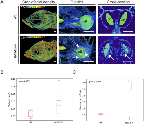
Abnormal mineralisation of amorphous material within the adult inner ears and higher BMD in ncoa3−/−. A) 3D renders from μCT images of wt and ncoa3−/− of same age (1 year old). The head was color-coded to show bone mineral density (g.cm3HA; min = 0.338; max = 1.124). Note that craniofacial bones in ncoa3 mutants have higher density compared to wt. Otoliths (otl = arrows) were zoomed in. Abnormal mineralisation (dashed arrows) is observed attached to the otoliths. A cross section picture was taken to show the mineralised amorphous material (arrows) juxtaposed to the otoliths. B) Volume of otoliths. C) Bone mineral density of central region of otoliths. Non-parametric, two-tailed, independent Student’s t-Test was used as statistical analysis (p < 0.05). Scale bars = 500 μm.

Figure Data
Acknowledgments
This image is the copyrighted work of the attributed author or publisher, and ZFIN has permission only to display this image to its users. Additional permissions should be obtained from the applicable author or publisher of the image. Full text @ Hum. Mol. Genet.