FIGURE

Figure 8.

ID
ZDB-FIG-201121-164
Publication
Panlilio et al., 2020 - Developmental Neurotoxicity of the Harmful Algal Bloom Toxin Domoic Acid: Cellular and Molecular Mechanisms Underlying Altered Behavior in the Zebrafish Model
Other Figures
All Figure Page
Back to All Figure Page
Figure 8.

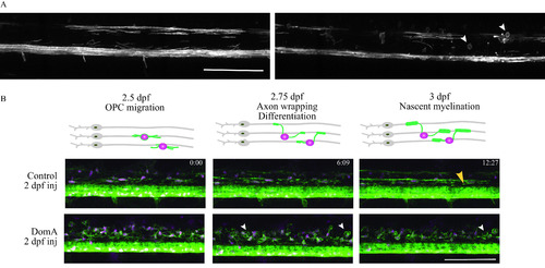
Time-lapse imaging of myelin sheath formation in zebrafish exposed to domoic acid (DomA) at 2 d postfertilization (dpf). (A) Tg(mbp:EGFP-CAAX) fish were used to visualize labeled myelin sheaths. Shown are representative images of DomA-exposed (0.14 ng) (right panel), and control larvae (left panel) that were exposed at 2 dpf and imaged at 3 dpf. White arrowheads denote the aberrant circular protrusions found in DomA-exposed larvae (control, n=5; DomA, n=10). (B) Stills from time-lapse imaging of a representative control and DomA-exposed transgenic fish [Tg(nkx2.2:mEGFP)×Tg(sox10:RFP)] from 2.5–3 dpf. Time stamps (hour:minutes) show the time elapsed. Diagrams above the images show the key developmental processes in the oligodendrocyte lineage during this time range (representative time-lapse image of control, n=5 and DomA, n=6). Large yellow arrowhead denotes an elongated myelin sheath. Small white arrowheads denote unusual circular myelin membranes. Scale bar: 100μm. Stills (B) were from a time lapse of control (Video S2) and DomA-exposed fish (Video S3).

Expression Data

Expression Detail
Antibody Labeling
Phenotype Data

Phenotype Detail
Acknowledgments
This image is the copyrighted work of the attributed author or publisher, and ZFIN has permission only to display this image to its users. Additional permissions should be obtained from the applicable author or publisher of the image. Full text @ Environ. Health Perspect.