Generation of <italic>smyhc1</italic> mutant lines and embryonic muscle development
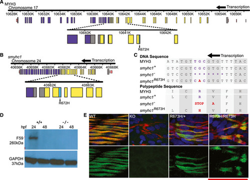
Schematic to scale of human MYH3 gene on chromosome 17. Noncoding regions are displayed in pink. Coiled coil domain (840–1,933 bp) displayed in purple. Motor domain (86–779 bp) displayed in yellow. Actin binding site (656–678/758–772 bp) shown in cyan. Location of R672H mutation is enlarged and labeled.
Schematic to scale of zebrafish smyhc1 gene on chromosome 24. Noncoding regions are displayed in pink. Coiled coil domain (842–1,929 bp) displayed in purple. Motor domain (85‐778 bp) displayed in yellow. Actin binding site (655–677 bp) displayed in cyan. Location of R672H mutation is enlarged and labeled.
Aligned DNA and amino acid sequences of MYH3 and smyhc1 alleles surrounding the smyhc1R673H and MYH3 R672H substitutions. The smyhc1− allele has a 7 base pair deletion that results in a frameshift in which one errant amino acid precedes a premature stop codon. The smyhc1R673H allele results from a G>A transition single point mutation.
Western blot of whole zebrafish larvae, Smyhc1 stained with the F59 antibody. smyhc1+/+ larvae express Smyhc1 at 24 hpf, but not at 48 hpf. smyhc1−/− larvae do not express Smyhc1.
Smyhc1 immunohistochemistry stain of 24 hpf (hour postfertilization) zebrafish larvae. Filamentous actin is stained with phalloidin (red). Nuclei are stained with DAPI (blue). Smyhc1 is stained with the F59 antibody (green) (Elworthy et al, 2008). smyhc1+/+, smyhc1R673H/+, and smyhc1R673H/R673H larvae display Smyhc1 in the muscle fibers at 24 hpf. smyhc1−/− larvae at 24 do not stain positively for Smyhc1. Scale bar represents 50 μm.
This image is the copyrighted work of the attributed author or publisher, and
ZFIN has permission only to display this image to its users.
Additional permissions should be obtained from the applicable author or publisher of the image.
Full text @ EMBO Mol. Med.
Your Input Welcome
Thank you for submitting comments. Your input has been emailed to ZFIN curators who may contact you if
additional information is required.
Oops. Something went wrong. Please try again later.