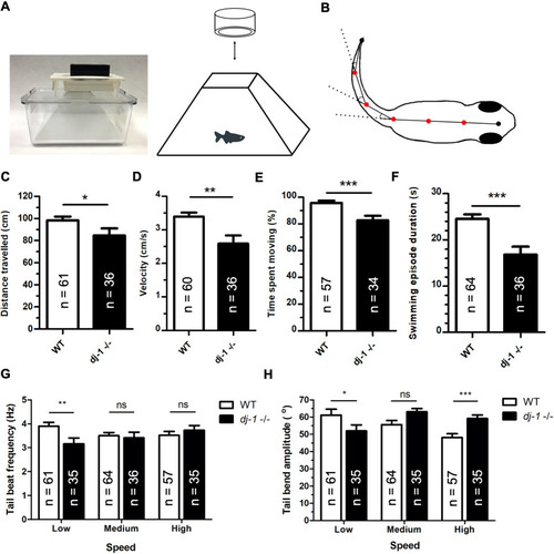
Analysis of extracted features reveals distinct movement in dj-1−/− zebrafish. (A) A photograph of the frustum insert designed to fit an Aquatics Habitat mating tank with a GoPro camera attached (left), providing a simple system for accurate, high-resolution video capture of adult zebrafish movement. A diagram of the fish inside the frustrum insert, recorded from above using the GoPro camera (right). (B) A diagram of the angles measured along the zebrafish trace, at the five vertices (red dots), when there is a bend in the tail. x and y coordinates were measured for the vertices and endpoints. Measurements were recorded at 100 frames/s from the video input, allowing analysis of selected features of movement. (C-H) The features of movement compared between dj-1−/− and wild type (WT) at 12 wpf including distance travelled (C), velocity (D), percentage of time spent moving (E), mean duration of a swimming episode (F), tail beat frequency at low, medium and high swimming speeds (G), and tail bend amplitude at low, medium and high swimming speeds (H) (single replicate). The number of replicates (n) is shown for each graph. Student’s t-tests (two-tailed, unpaired) were used to compare features that followed a normal distribution; the Mann–Whitney U-test (two-tailed) was used to compare non-parametric features. Data are mean± s.e.m., *P<0.05, **P<0.01, ***P<0.001; ns, not significant.
|
