
Fig. S2
- ID
- ZDB-FIG-200922-13
- Publication
- Gostic et al., 2019 - The dyslexia susceptibility KIAA0319 gene shows a specific expression pattern during zebrafish development supporting a role beyond neuronal migration
- Other Figures
- All Figure Page
- Back to All Figure Page
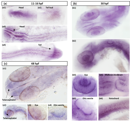
Supplementary Figure S2. In situ hybridization suggests a specific spatiotemporal expression pattern for kiaa0319. (a) Expression up to 16 hpf. At the 3 somite stage, kiaa0319 is expressed throughout the embryo with the strongest signal in the head (a1) and to a lesser extent, in the tail bud (a2). At the 14 the somite stage, high expression continues in the head (a3) and is visible along the developing body midline and in the tail (a3) (a4). All images are dorsal views with the head on the left side. At 30 hpf (b) kiaa0319 is still expressed throughout the embryo but strong expression emerges in specific structures observed from dorsal (b1) and lateral (b2) views. Details of expression are shown for the eyes (b3; dorsolateral view), the midbrain-hindbrain boundary (b4; dorsal view), the otic vesicles (b5; dorsolateral view) and the notochord (b6; lateral view). In a dorsal view at 48 hpf (c), kiaa0319 expression in the eyes is diminished, while signal in the telencephalon emerges (c1). The signal in the telencephalon is particularly visible in a lateral view (c2) along with expression in the eyes and the region around the notochord. Detailed dorsolateral views of the eye (c3) and otic vesicle (c4) show weaker intensity at these structures when compared to the pattern observed at 30 hpf. |