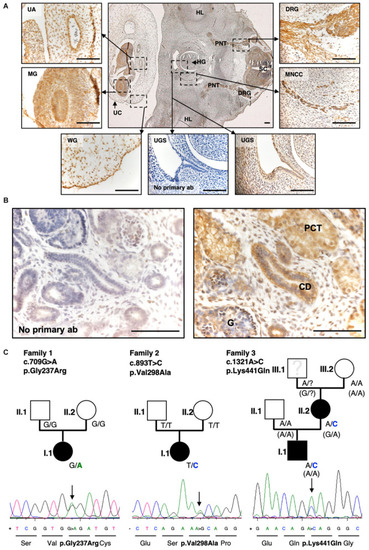
Exome sequencing and targeted Resequencing in families with BEEC phenotype identified disease variants in SLC20A1.(A) IHC of a transverse section of a 6-week-old human embryo. SLC20A1 was immunodetected (brown) in the urogenital sinus (UGS), which develops into the urinary bladder. Furthermore, (textitSLC20A1 was also detected in the spinal cord (SC), dorsal root ganglia (DRG), peripheric nerve trunk (PNT), hind limb (HL), hindgut (HG), migrating neural crest cells (MNCC), umbilical cord (UC), Wharton’s jelly (WG), midgut (MG), and umbilical artery (UA). Scale bars = 100 μm. (B) Histology section of the human 10-week-gestation metanephric metanephros. Note prominent SLC20A1 immunostaining (brown) in the proximal tubule (PT) and the collecting duct (CD). The glomerulus (G) shows a fainter signal. Scale bars: 100 μm. (C) Exome sequencing of eight CE case-parent-trios revealed de novo variant c.709G > A (p.Gly237Arg) in SLC20A1 in family 1 (Reutter et al., 2016). Resequencing of 690 individuals with BEEC led to identification of two more variants in individuals with CBE: c.893T > C (p.Val298Ala) in family 2 as de novo change c.1321A > C (p.Lys441Gln) with maternal inheritance (maternal phenotype: fusion defect of pelvic bone, mild phenotype) in family 3. Pedigrees of all three families are shown with genotypes of all individuals indicated. In family 3, the maternal grandfather (Figure 1B, family 3, person III.1) was not available for testing. For haplotype analysis of all available family members (Figure 1B, family 3, III.2, II.1, II.2, I.1), we used the synonymous marker rs4849091 at chromosomal position chr2:113404708 A > G (p.Leu101=) of the canonical transcript ENST00000272542.7. Genotypes of rs4849091 are shown in brackets. All variants are heterozygote changes and result in missense variants as shown in Sanger sequences including amino acid sequences below each pedigree.)
|
