|
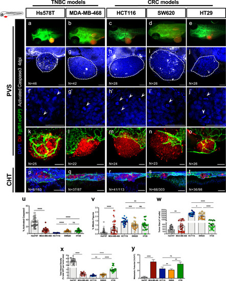
Characterization of zebrafish TNBC and CRC xenografts models.Human cancer cell lines (Hs578T, MDA-MB-468, HCT116, SW620 or HT29) were fluorescently labeled with DiI (red) and injected into the perivitelline space (PVS) of 2 days post fertilization (dpf) Tg(fli1:eGFP) zebrafish larvae (a–e). At 4 days post injection (dpi), zebrafish xenografts were evaluated regarding: apoptotic index—% of activated Caspase3 (f–j, u), mitotic index—% mitotic figures (f’–j’, v), tumor size (f–j, w), angiogenic capacity (k–o, x) and metastatic potential (p–t, y). White arrowheads indicate mitotic figures. Apoptotic index (u, ****P < 0.0001), mitotic figures (v, Hs578T versus MDA-MB-468 ****P < 0.0001, ***P = 0.0002, **P = 0.0049), tumor size (w, ****P < 0.0001, **P = 0.0065, *P = 0.0129), total vessel density (x, ****P < 0.0001, **P = 0. 0045) and metastatic potential (y, ****P < 0.0001, HCT116 versus SW620 **P = 0.0036, SW620 versus HT29 **P = 0.0048, Fisher’s exact test) are expressed as AVG ± SEM. The number of xenografts analyzed are indicated in the representative images and each dot represents one zebrafish xenograft. Results are from 3 (Hs578T and MDA-MB-468) and 2 (HCT116, SW620 and HT29) independent experiments, which are highlighted in different colors corresponding to each individual experiment. Statistical analysis was performed using an unpaired t-test or Fisher’s exact test. Statistical results: (ns) > 0.05, *P ≤ 0.05, **P ≤ 0.01, ***P ≤ 0.001, ****P ≤ 0.0001. Scale bars represent 50 μm. All images are anterior to the left, posterior to right, dorsal up and ventral down.
|