|
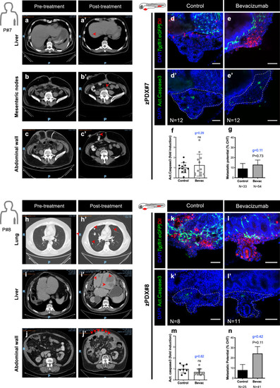
zPDX bevacizumab treatment response may predict relapse and correlates with patients' outcome.Computerized tomography scans of Patient#7 (a–c’) and Patient#8 (h–j’) of different regions, including liver (a, a’, i, i’), lung (h, h’), mesenteric region (b, b’) and abdominal wall (c, c’, j, j’) in both pre- and post-treatment settings. Red arrows highlight metastasis. Human patient surgical/biopsy samples were injected into the PVS of 2 dpf Tg(fli1:eGFP) zebrafish larvae. zPDXs were treated in vivo with bevacizumab and compared with untreated controls. At 4 dpi, zebrafish xenografts were imaged by confocal microscopy (d–e’, k–l’). Activated caspase3 was quantified in both groups (f, zPDX#7 P = 0.4682 and g = 0.29; m, zPDX#8 P = 0.1817 and g = 0.82). In parallel, the percentage of xenografts that display micrometastasis in the CHT region was quantified (g, zPDX#7 P = 0.73 and g = 0.11; n, zPDX#8 P = 0.11 and g = 0.42). The outcomes are expressed as AVG ± SEM. Number of zPDX analyzed for each condition is indicated in the figure. Results are from 1 independent experiment. Statistical analysis was performed using an unpaired t-test for apoptosis and a Fisher’s exact test for micrometastasis. Statistical results: (ns) > 0.05, *P ≤ 0.05, **P ≤ 0.01, ***P ≤ 0.001, ****P ≤ 0.0001. Cohen’s D 1988 scale of effect size with Hedges’ g correction (g): g = 0,2 low; g = 0.5 moderate; g = 0.8 high. Scale bars represent 50 μm. All images are anterior to the left, posterior to right, dorsal up, and ventral down.
|