
Fig. s2
- ID
- ZDB-FIG-200618-7
- Publication
- Xie et al., 2019 - Schwann cell precursors contribute to skeletal formation during embryonic development in mice and zebrafish
- Other Figures
- All Figure Page
- Back to All Figure Page
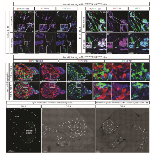
Leakage control in Plp1CreERT2 mouse embryos (A-D) Genetic tracing of Plp1CreERT2;R26RYFP/+ embryos from E11.5 till E12.5 (A-B) or E17.5 (C-D) revealed 12.33% of the P0+ Schwann cells along the TUJ1+ nerves labeled genetically at E13.5, but the percentage increased to 56.56% at E17.5. (E-F) Plp1CreERT2;R26RYFP/+ did not genetically label tyrosine hydroxylase (TH)-positive neurons of the sympathetic ganglia (neural crest origin) when traced from E11.5 to E17.5 (E) or E12.5 to E17.5 (F). Neurofilaments were visualized with an antibody against NF200. (G) A representative scan of an entire E11.5 Plp1CreERT2;R26RYFP/+ embryo not injected with tamoxifen, shows no YFP signal other than auto-fluorescence from the internal organs and blood vessels. The laser power and digital gain were set at much higher levels (50%) than routine (around 6.5%) for visualization purposes . The white dashed lines outline the embryo. (H-I) Representative scans of sagittal facial sections of E17.5 Plp1CreERT2;R26RYFP/+ embryos either not injected with tamoxifen (H) or injected with corn oil at E11.5 (I), show no sporadic recombination in the absence of tamoxifen. The white dashed lines outline cartilage elements. |