
Fig. s8
- ID
- ZDB-FIG-200618-13
- Publication
- Xie et al., 2019 - Schwann cell precursors contribute to skeletal formation during embryonic development in mice and zebrafish
- Other Figures
- All Figure Page
- Back to All Figure Page
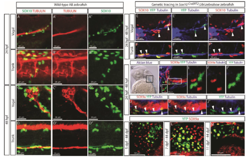
(A-D) Cells immuno-stained positively for SOX10 (green) were seen to be located along tubulin (red)-positive nerves in both the vagal (A, C) and trunk (B, D) regions of wild-type AB zebrafish at 24 (A-B) and 48 (C-D) hpf. (E-F) Genetic tracing in Sox10CreERT2;Ubi:zebrabow zebrafish larva from 24 to 48 hpf revealed overlap between genetically labeled YFP+ cells (green) and SOX10+ cells (red) along the Tubulin+ nerves (blue) in both the vagal (E) and trunk (F) nerve regions. Arrowheads in (E-F) indicate examples of YFP and SOX10 double-positive cells. (G) Whole-mount skeletal preparation revealed appearance of the first alcian blue-positive condensation at the region of otic vesicle at 48 hpf and this area was further examined in Sox10CreERT2;Ubi:zebrabow zebrafish larva genetically traced from 24 to 48 hpf (H-J). Few YFP+ cells detached from the nerve were observed at this time-point, but those were not SOX9a+ (I), at the same time YFP+ and SOX9a+ double positive cells (highlighted by the white arrowheads) were observed tightly associated with the Tubulin+ nerve (J). (K-M) Genetic tracing in Sox10CreERT2;Ubi:zebrabow zebrafish after formation of SOX10+ cartilage elements, i.e., from 5 (K), 7 (L) or 11 dpf (M) until 44 dpf revealed a pattern of labeling that differed from that of SCPs labeled at 24 or 48 hpf (Fig. 4C-J). Images in A-F are single confocal slices of the stained samples. |