
Fig. s7
- ID
- ZDB-FIG-200618-12
- Publication
- Xie et al., 2019 - Schwann cell precursors contribute to skeletal formation during embryonic development in mice and zebrafish
- Other Figures
- All Figure Page
- Back to All Figure Page
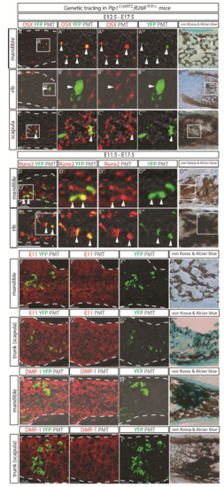
(A-C) In Plp1CreERT2;R26RYFP/+ embryos traced from E12.5 to E17.5 SCPs descendants were detected positive for osterix (OSX) marker for osteo-progenitors in the mandible (A), ribs (B) and scapula (C). Traced cells and OSX+ cells were visualized with an anti-GFP antibody (green) and anti-Osx antibody (red), respectively. The white arrowheads point to the double-positive cells. (D-E) The Plp1CreERT2;R26RYFP/+ embryos traced from E11.5 to E17.5 were stained with an anti-GFP antibody (green) and antibody against a pre-osteoblast marker RUNX2. (F-I) Domains enriched in osteocytes in the mandible (F, H) and scapula (G, I) in Plp1CreERT2;R26RYFP/+ embryos traced from E11.5 to E17.5 were visualized with antibodies against E11/gp38 osteocyte marker (F-G) or dentin matrix protein (DMP1) (H-I). Panels A""-E"" and F"'-I"' depict the same tissue sections as panels A-I, but stained with von Kossa & Alcian blue following completion of confocal scans. The white dashed lines outline the mineralized portion of the bone as indicated by the von Kossa & Alcian blue staining. |