|
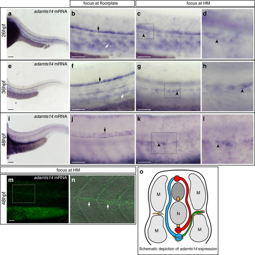
<italic>adamts14</italic> expression can be detected in the floorplate and at the horizontal myoseptum.a–l Whole mount in situ hybridization for adamts14. a–d At 26 hpf, adamts14 transcripts are detectable in the floorplate (black arrow), in cells along the ventral aspect of the segment boundaries (white arrow) and in cells at the HM (black arrowhead). d Zoom-in of the squared region in c. e–h At 36 hpf, adamts14 expression is visible in the floorplate (black arrow), in cells located at the level of the axial blood vessels (white arrow) and in cells at the HM (black arrowhead). h Zoom-in of the indicated region in g. i–l Expression of adamts14 is still apparent in the floorplate (black arrow) at 48 hpf as well as in cells at the HM (black arrowhead). l Enlargement of the boxed region in k. m, nadamts14 RNA granules can be detected in cells located in the HM region by RNAscope at 48 hpf. n Overlay of the bright field and confocal image for the indicated region in m, showing adamts14 RNA granules in green. o Schematic cross view of the trunk illustrating the adamts14 expression domains (yellow). Scale bars in a, e, i: 100 µm; b, c, f, g, j, k, m: 50 µm. DA dorsal aorta, hpf hours post fertilization, HM horizontal myoseptum, M muscle, N notochord, PCV posterior cardinal vein, PL parachordal lymphangioblast.
|