|
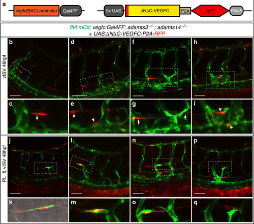
Local Vegfc processing at the HM is required to guide endothelial cells to this region.a Schematic depiction of the mature human VEGFC overexpression construct and the vegfc:Gal4FF transgenic line, which was used to drive mosaic expression of the construct upon transient injection. b–qvegfc:Gal4FF positive embryos were injected with the overexpression construct at the one-cell stage and imaged at 48 hpf. All embryos shown are adamts3; adamts14 double mutant, transgenic for flt4:mCitrine (green) and express UAS:ΔNΔC-VEGFC-P2A-RFP (red). b, c Expression of UAS:ΔNΔC-VEGFC-P2A-RFP in hypochord cells (white arrow) rescues vein formation. d, e Local vISV rescue upon expression of mature VEGFC in DA cells (white arrowhead). f–i Venous hyper-sprouting with ectopic venous structures formed in the vicinity of hypochord cells (f, g, arrows) or DA cells (h, i, arrowheads) with expression of the ΔNΔC-VEGFC construct. j, kUAS:ΔNΔC-VEGFC-P2A-RFP positive fibroblasts recruit PL cells to the HM region. l, m Expression of mature VEGFC in hypochord cells and in fibroblasts at the HM rescues vISV and PL formation in adamts3; adamts14 double mutant embryos. Note the close proximity of the PL cell to the ΔNΔC-VEGFC producing fibroblasts. n, o Combined expression of mature VEGFC in DA cells and fibroblasts at the HM results in the formation of vISV and PL cells close by. p, q Simultaneous expression of ΔNΔC-VEGFC in hypochord cells and in fibroblasts at the HM results in a local rescue of PL and vISV formation but can additionally lead to a hyper-branching of venous structures at the level of the DA. c, e, g, i, k, m, o, q: Zoom-in image of the indicated regions in b, d, f, h, j, l, n, p. Scale bars: 50 µm. DA dorsal aorta, HM horizontal myoseptum, hpf hours post fertilization, PL parachordal lymphangioblast, vISV venous intersegmental vessel.
|