
Fig. 1
- ID
- ZDB-FIG-200615-32
- Publication
- Blume et al., 2020 - Microglia in the developing retina couple phagocytosis with the progression of apoptosis via P2RY12 signaling
- Other Figures
- All Figure Page
- Back to All Figure Page
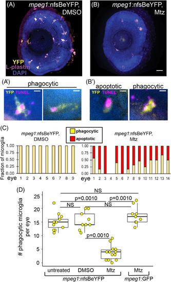
Metronidazole (Mtz)‐mediated depletion of microglia in the retina of mpeg1 :nfsBeYFP zebrafish embryos during development. (A, B), Images of whole eyes from mpeg1 :nfsBeYFP embryos following immersion in DMSO, A, or Mtz, B, from 24 to 72 hpf, stained for L‐plastin (purple) and DAPI (blue) with YFP expression also visualized (yellow). All L‐plastin+ cells in the developing retina express YFP, A, and these cells are reduced upon Mtz treatment, B. Scale bar in B = 20 μm and applies to A and B. A′. Examples of YFP+ microglia in control retinas with phagocytic morphology engulfing TUNEL+ dying cells (magenta). B′. Examples of morphology indicating apoptotic TUNEL+YFP+ microglia in retinas from mpeg1 :nfsBeYFP retinas treated with Mtz (left) and remaining phagocytic YFP+ microglia engulfing TUNEL+ puncta (right). Scale bars in A′ and B′ = 5 μm. C, YFP+ microglia in mpeg1 :nfsBeYFP retinas treated with DMSO (left) or Mtz (right) were assigned phagocytic or apoptotic morphology. Graphs show the fraction of microglia with each morphology in each eye examined. D, To determine the effective depletion, the number of phagocytic microglia in the retina in each indicated group was quantified. Box plots are shown for each group with each individual data point shown in yellow circles. A one‐way analysis of variance (ANOVA) (P = 2.04 × 10⁻¹⁴) was performed, followed by Tukey's HSD post hoc test. P values shown for pairwise comparison with P < .05; NS = not significant. Phagocytic microglia were depleted only in mpeg1 :nfsBeYFP embryos treated with Mtz. Samples sizes: mpeg1 :nfsBeYFP untreated = 13 eyes from 13 fish, mpeg1 :nfsBeYFP DMSO treated = 9 eyes from 5 fish, mpeg1 :nfsBeYFP Mtz treated = 14 eyes from 10 fish, mpeg1 :GFP Mtz treated = 9 eyes from 6 fish |