
Fig. 5
- ID
- ZDB-FIG-200615-28
- Publication
- Blume et al., 2020 - Microglia in the developing retina couple phagocytosis with the progression of apoptosis via P2RY12 signaling
- Other Figures
- All Figure Page
- Back to All Figure Page
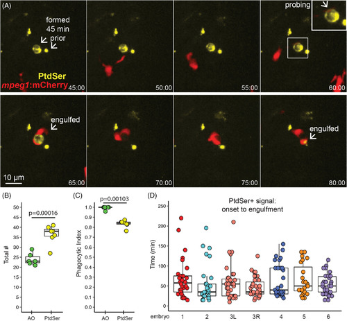
Microglial sensing of apoptotic neurons displaying Phosphatidyl Serine. A, Real‐time imaging of microglia (red) sensing retinal neurons with exposed phosphatidyl serine (PtdSer, yellow). Selected panels are shown for an imaging session. Timestamp (hour:minute, bottom right) is relative to YFP onset in the selection. In contrast to AO, YFP+ cell bodies were present (here, for ~1 hour) prior to microglial sensing, probing, and engulfment. Scale bar applies to all images. Inset (upper right panel) shows enlargement of the PtdSer+ cell probed by a microglial process. B, Box plots show total number of PtdSer+ cells (n = 6 embryos) over the imaging session, compared to AO+ numbers (n = 6 embryos). Individual circles overlaid on box plots represent each sample. P value (Student's 2‐tailed t test) is shown. C, Phagocytic index of apoptotic cells detected by AO or PtdSer. The phagocytic index is defined as the fraction of cells observed to be cleared by microglia during the imaging session. Box plots, with individual measurements overlaid (circles), are shown. P value from the generalized liner model with binomial family is shown. D, For each individual PtdSer+ cell counted, the time of PtdSer exposure from onset to microglial engulfment was calculated. Each box plot represents one eye from one embryo, except for the salmon colored plots, which show two eyes from a single embryo. Each circle overlaid on the box plot represents one PtdSer+ cell |