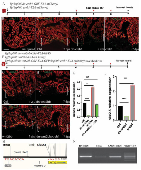
Wnt2bb/c-jun/creb pathway mediated zebrafish heart regeneration through nkx2.5. (A,F) Schematic of the experimental plan after apex amputation. (B) In uninjured hearts, nkx2.5 was detectable in cardiomyocyte nuclei throughout the myocardium. (C) Following ventricular resection, nkx2.5 was induced at the apical cell edge of wounded myocardia at 7 dpa. (D)Nkx2.5 was inhibited in dn-creb hearts following ventricular resection. (E)Nkx2.5 was enhanced in creb-ORF-overexpressing hearts following ventricular resection at the apical cell edge of wounded CMs. (G) Confocal image analyses displaying nkx2.5 at wounded myocardial cell edges in control hearts (I), elevated nkx2.5 in injured Tg(hsp70l:wnt2bb) hearts (H), diminished nkx2.5 in Tg(hsp70l:dn-wnt2bb) hearts (J) and rescued in Tg(hsp70l:dn-wnt2bb;hsp70l:creb1) hearts. (M,N) Schematic of the CREB binding site CRE (TGACATCA) at the upstream 3.7 kb of nkx2.5 ATG (I) and CHIP experiment show that creb can bind the CRE site of Nkx2.5. (K,L) Bar charts showing the quantification of relative nkx2.5 expression in heat-shocked, injured wild-type (ctrl), Tg(hsp70l:dn-wnt2bb), Tg(hsp70l:wnt2bb), Tg(hsp70l:creb1), Tg(hsp70l:dn-wnt2bb), and Tg(hsp70l:dn-wnt2bb;hsp70l:creb) fish hearts. The data from five hearts in each group are presented. Error bars: ± 1 SD. Significance was determined using a Student’s t-test: **P < 0.01, ***P < 0.001, ****P < 0.0001. Green, nkx2.5; Red, MF20; Brackets, amputation area; Scale bar, 100 μm. Fluorescent intensities were measured at the injury border zone using ImageJ.
|
