FIGURE

Figure 8

ID
ZDB-FIG-200608-8
Publication
Albadri et al., 2020 - Expression of a Barhl1a reporter in subsets of retinal ganglion cells and commissural neurons of the developing zebrafish brain
Other Figures
All Figure Page
Back to All Figure Page
Figure 8

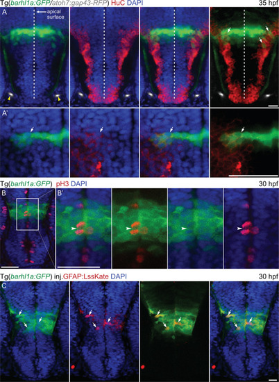
Barhl1a:GFP in the preoptic diencephalon marks populations of mitotic progenitors and differentiating neurons. Images represent single confocal Z-stacks through the diencephalon of Tg(barhl1a:GFP;atoh7:gap43-RFP) embryos at 35 hpf (A, A’) and 30 hpf (B, C). Frontal view, dorsal is up. Embryos in (A-B’) have been immunohistochemically stained with anti-GFP, anti-HuC (A, A’) and anti-pH3 (B, B’) antibodies (in red) as well as counterstained with DAPI (in blue). (A’) magnifications of the images in (A) in correspondence with the barhl1a:GFP expressing cell bodies. (A) yellow arrowheads point at the optic nerves from each side marked by Atoh7:gap43-RFP (in gray). White arrows point at Barhl1a:GFP cells co-labelled with HuC, a marker of differentiating neurons located in the basal half of the neuroepithelium. In (B) and its magnification (B’), Barhl1a:GFP cells are also co-labelled by pH3, which marks mitotic neurons located at the apical surface of the neuroepithelium (white arrowhead in B’). (C) 30 hpf Tg(barhl1a:GFP) embryo injected with the gfap:lssKate DNA construct shows sparse LssKate labelling in the diencephalon (in red); which co-localize with GFP (green). Scale bars: (A–C) 50 µm, (A’,B’) 20 µm.

Expression Data

Expression Detail
Antibody Labeling
Phenotype Data

Phenotype Detail
Acknowledgments
This image is the copyrighted work of the attributed author or publisher, and ZFIN has permission only to display this image to its users. Additional permissions should be obtained from the applicable author or publisher of the image. Full text @ Sci. Rep.