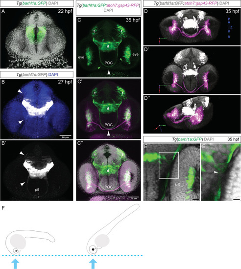
Developmental dynamic of Barhl1a:GFP cells in the diencephalon. Images represent projections of Z-stack (A–C”, E, E’) and 3D reconstructions (D–D”) of the anterior brain of fixed embryos at 22 (A), 27 (B-B’) and 35 hpf (C-C”, D-D” and E-E’) counterstained with DAPI. Images are in frontal view, dorsal is to the top. (A) Frontal view of the anterior brain of a Tg(barhl1a:GFP) transgenic embryo at 22 hpf showing Barhl1a:GFP (in green) in the presumptive diencephalon. DAPI staining is shown in gray. (B-B’) Frontal view of a Tg(barhl1a:GFP) transgenic embryo at 27 hpf. DAPI in (B) is shown in blue. White arrowheads point at Barhl1a:GFP projections (in gray) that extend and cross the midline ventrally and dorsally, respectively. The fibers that cross ventrally form a bundle of fibers along the presumptive optic tract. At this stage, few GFP-positive cells likely corresponding to pituitary cells can also be detected (pit, B’). (C-C”) Frontal view of an 35 hpf Tg(barhl1a:GFP;atoh7:gap43-RFP) double transgenic embryo. DAPI staining is shown in gray in C’. Barhl1a:GFP cells (in green) can be seen in tectal (t) and diencephalic (d) domains as well as in the eye. Bundles of Barhl1a:GFP fibers can be clearly seen crossing the ventral midline along the post optic commissure (POC). At this stage, the Atoh7:gap43-RFP positive RGCs (magenta) extend their axons out of the retina to form the optic nerve. The optic nerves cross contralaterally at the optic chiasm (white arrowhead) in close proximity to the diencephalic GFP-positive fibers forming the POC. The green arrowheads in (C) point at the few Barhl1a:GFP RGC fibers (in green), which are contained within the big bundle of magenta RGC fibers shown in C’ and C”. (D-D”) 3D reconstruction and turnaround of the anterior head region of a double transgenic Tg(barhl1a:GFP;atoh7:gap43-RFP) embryo at 35 hpf highlighting the optic nerves (in magenta) alongside the Barhl1a:GFP fibers (in gray). DAPI staining is in gray. In all three panels the view is from the posterior side of the stack/embryo; P, posterior; A, anterior; Z, z-axis. (E-E’) Enlarged confocal image (E) and its magnification (E’) of a frontal view of an Tg(barhl1a:GFP) transgenic embryo highlighting the fibers originating from Barhl1a:GFP cells located in the diencephalon (arrowheads). (F) Schematic cartoons showing approximate embryo orientation and plane of confocal imaging. Pit, pituitary. Scale bars: (A) 28 µm, (B-B’) 66 µm, (C-C”) 70 µm, (E) 20 µm, (E’) 50 µm.
|
