FIGURE

Figure 1

ID
ZDB-FIG-200608-1
Publication
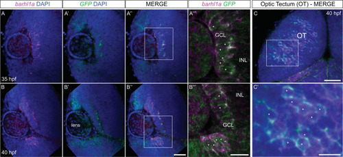
Albadri et al., 2020 - Expression of a Barhl1a reporter in subsets of retinal ganglion cells and commissural neurons of the developing zebrafish brain
Other Figures
All Figure Page
Back to All Figure Page
Figure 1

Expression of endogenous barhl1a is reflected by barhl1a:GFP in the retina and brain. (A–B”’) Confocal optical section in frontal view (dorsal is to the top) through the retina of a 35 hpf (A - A’’’) and 40 hpf (B - B’’’) embryo after FISH against endogenous barhl1a (A, B in magenta) and the GFP transgene (A’, B’ in green). Retinae are counterstained with DAPI (blue). (A”, B”) merging of the two channels shows co-localization of the two transcripts in the GCL. (A”’, B”’) magnifications of (A”) and (B’’) (white squares), respectively, without the DAPI channel. (C-C’) Confocal imaging into the tectum of a 40 hpf embryo after FISH and DAPI conterstaining shows co-localization of barhl1a with GFP. Images represent a Z-stack in dorsal view (anterior is to the top) into the optic tectum (OT). (C’) magnification of (C) (white square). Asterisks highlight regions with colocalizing signals. GCL: ganglion cell layer, INL: inner nuclear layer, OT: optic tectum. Scale bars: (A-A”, B-B”, C) 50 µm, (A’’’, B’’’, C’) 20 µm.

Expression Data

Expression Detail
Antibody Labeling
Phenotype Data

Phenotype Detail
Acknowledgments
This image is the copyrighted work of the attributed author or publisher, and ZFIN has permission only to display this image to its users. Additional permissions should be obtained from the applicable author or publisher of the image. Full text @ Sci. Rep.