FIGURE

Fig. S2

ID
ZDB-FIG-200514-51
Publication
Weber et al., 2020 - Zebrafish disease model of human RNASET2 deficient cystic leukoencephalopathy displays abnormalities in early microglia
Other Figures
All Figure Page
Back to All Figure Page
Fig. S2

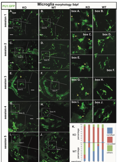
Microglia morphology of RNASET2-deficient embryos and larvae in independent clutches.
Zebrafish of the transgenic reporter strain Tg:(PU1:GFP) were analyzed by in vivo confocal laser scanning microscopy (CLSM). Data shown in Figure 1 were filtered to show the morphology of mutant and WT larvae from 5 independent clutches (session 1-5).
A-J Projections of confocal z-stacks through the brain showing microglia (green, arrows). Box A.-J (right panels) showing ramified and vacuoled microglia at increased size as in the respective images A.-J. (left panels). Mutant and WT larvae from the same session are opposed (Session 1A. mutant & B. WT; Session 2C. mutant & D. WT; Session 3E. mutant F. WT; Session 4G. mutant & H. WT; Session 5I. mutant & J. WT
K: Depiction of the morphological categories (ramified, vacuoled, others) of microglia at 5 dpf from 5 sessions.

Expression Data

Expression Detail
Antibody Labeling
Phenotype Data

Phenotype Detail
Acknowledgments
This image is the copyrighted work of the attributed author or publisher, and ZFIN has permission only to display this image to its users. Additional permissions should be obtained from the applicable author or publisher of the image. Full text @ Biol. Open