FIGURE

Fig. S1

ID
ZDB-FIG-200514-50
Publication
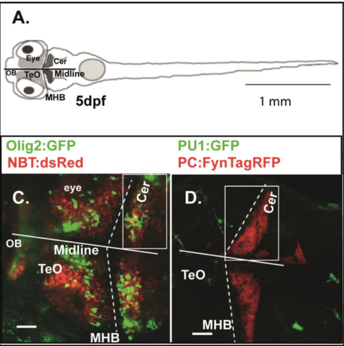
Weber et al., 2020 - Zebrafish disease model of human RNASET2 deficient cystic leukoencephalopathy displays abnormalities in early microglia
Other Figures
All Figure Page
Back to All Figure Page
Fig. S1

Transgenic zebrafish lines and analyzed zebrafish brain regions
A: 
Cartoon of a zebrafish larvae on 5 dpf showing the regions analyzed in this study. TeO Tectum opticum; OB Olfactory bulb; Cer Cerebellum; MHB Midbrain-hindbrain boundary. B: WT zebrafish larvae of compound transgenics Tg:(NBT:dsRed)/Tg:(Olig2:GFP) analyzed by in vivo CLSM. Projection of a confocal z- stack highlighting neurons (red) and oligodendrocyte precursor cells (green) on 5 dpf. C: WT zebrafish larvae of compound transgenics Tg:(PU1:GFP)/ Tg:(PC:FynTagRFP) analyzed by in vivo CLSM. Projection of a confocal z-stack highlighting microglia (green) and purkinje cells (red) on 5 dpf. Size markers 40 μM.

Expression Data

Expression Detail
Antibody Labeling
Phenotype Data

Phenotype Detail
Acknowledgments
This image is the copyrighted work of the attributed author or publisher, and ZFIN has permission only to display this image to its users. Additional permissions should be obtained from the applicable author or publisher of the image. Full text @ Biol. Open