
|
Fig. S2
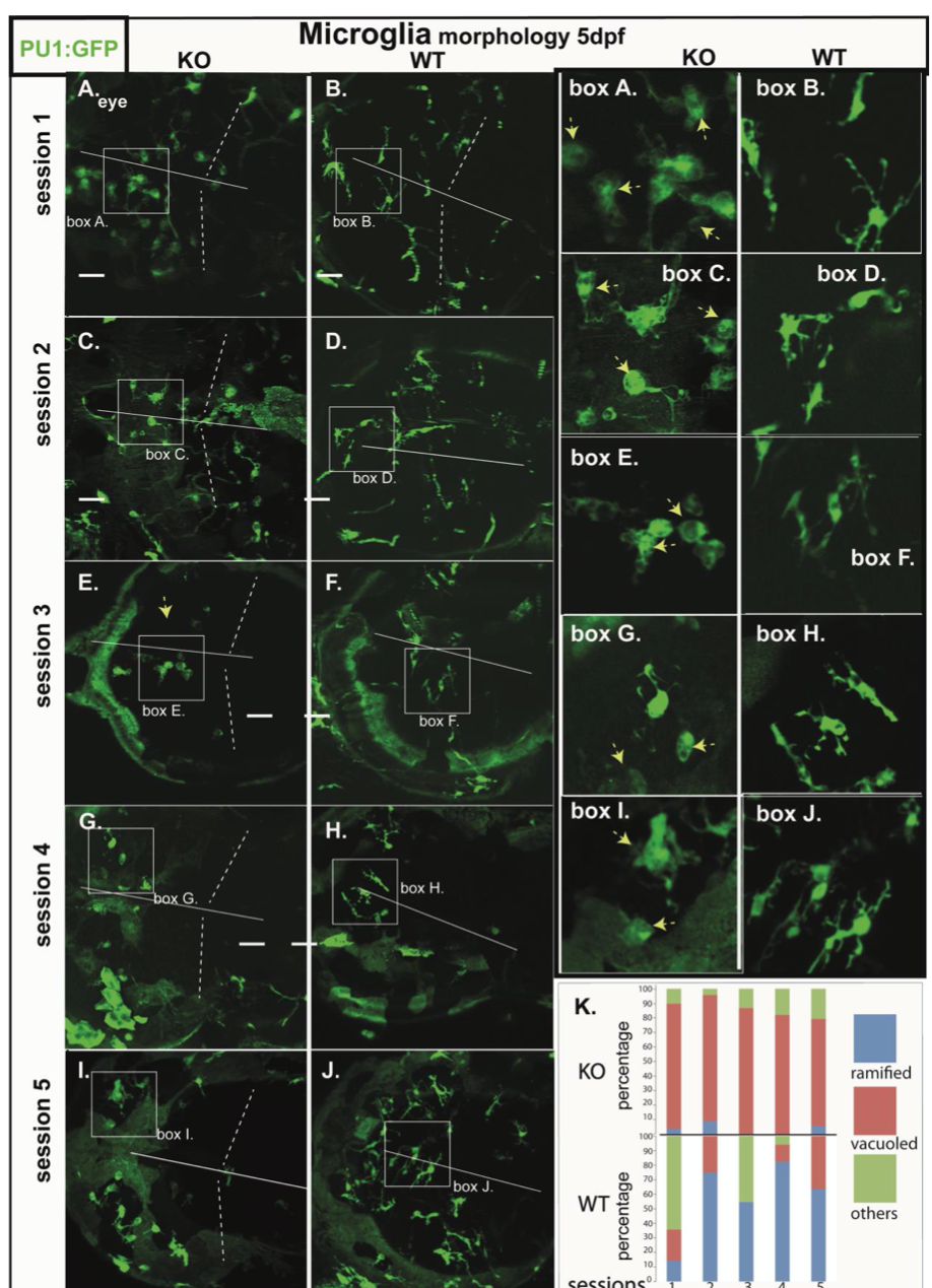
Microglia morphology of RNASET2-deficient embryos and larvae in independent clutches.
Zebrafish of the transgenic reporter strain Tg:(PU1:GFP) were analyzed by in vivo confocal laser scanning microscopy (CLSM). Data shown in Figure 1 were filtered to show the morphology of mutant and WT larvae from 5 independent clutches (session 1-5).
A-J Projections of confocal z-stacks through the brain showing microglia (green, arrows). Box A.-J (right panels) showing ramified and vacuoled microglia at increased size as in the respective images A.-J. (left panels). Mutant and WT larvae from the same session are opposed (Session 1: A. mutant & B. WT; Session 2: C. mutant & D. WT; Session 3: E. mutant & F. WT; Session 4: G. mutant & H. WT; Session 5: I. mutant & J. WT
K: Depiction of the morphological categories (ramified, vacuoled, others) of microglia at 5 dpf from 5 sessions.