FIGURE

Figure 5

ID
ZDB-FIG-200423-70
Publication
Cozzolino et al., 2020 - Evolution of Epileptiform Activity in Zebrafish by Statistical-Based Integration of Electrophysiology and 2-Photon Ca2+ Imaging
Other Figures
All Figure Page
Back to All Figure Page
Figure 5

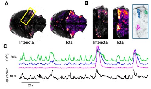
Identification of the micro-circuitry associated with different epileptic-like events. (A) Cross-correlation maps computed during two different intervals in th presence of only interictal activity (left) or during large ictal events (right). The cross-correlation maps have been normalized to the respective maximum value. (B) Details of the Cb, as outlined by the yellow rectangle (300 μm high) in panel A, showing the different sources of LFP signal during these two different classes of events. The finder map on the right shows three areas belonging to the cerebellum that are differentially active during interictal and ictal-like events. (C) ΔF/F0 measured in the three areas outlined by the corresponding colors in B. While all areas respond to the three large ictal events, the neuronal territory involved in interictal activity (green area and trace) is far more limited.

Expression Data

Expression Detail
Antibody Labeling
Phenotype Data

Phenotype Detail
Acknowledgments
This image is the copyrighted work of the attributed author or publisher, and ZFIN has permission only to display this image to its users. Additional permissions should be obtained from the applicable author or publisher of the image. Full text @ Cells