FIGURE

Figure 2

ID
ZDB-FIG-200423-67
Publication
Cozzolino et al., 2020 - Evolution of Epileptiform Activity in Zebrafish by Statistical-Based Integration of Electrophysiology and 2-Photon Ca2+ Imaging
Other Figures
All Figure Page
Back to All Figure Page
Figure 2

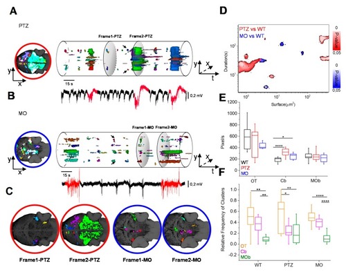
Dynamic evolution of the Ca2+ domains. (A,B) The volume representation depicts the active pixels from the binary stacks, clustered in domains of different colors of PTZ and MO larvae. The frame on the left is the projection of active domains during the entire period. The LFP events in red have high Ca2+ recruitment (>700 voxels). (C) The shaded sections in A and B are the spatial patterns of activation during the interictal-like events (frame 1) and the generalized events (frame 2) in the PTZ (red) and MO (blue). (D) Difference of the spatio-temporal morphology of PTZ and MO clusters with respect to WT clusters. The maximum surface occupied by Ca2+ clusters is plotted in relation to their duration. The color bars represent the p-value of the difference with the WT model. We report only the significative differences (p-value ≤ 0.05). 4 recordings for each larva, n = 3 larvae for each condition. (E) Comparison of the amounts of detected voxels for the three models in different anatomical regions. (F) Anatomical localization of the origin of the recruited neuronal clusters with volume > 300 voxels. The data have been normalized to the total number of active domains measured in each recording session (* p ≤ 0.05, ** p ≤ 0.01, *** p ≤ 0.001, **** p ≤ 0.0001).

Expression Data

Expression Detail
Antibody Labeling
Phenotype Data
Fish:
Condition:
Knockdown Reagent:
Observed In:
Stage: Day 5

Phenotype Detail
Acknowledgments
This image is the copyrighted work of the attributed author or publisher, and ZFIN has permission only to display this image to its users. Additional permissions should be obtained from the applicable author or publisher of the image. Full text @ Cells