
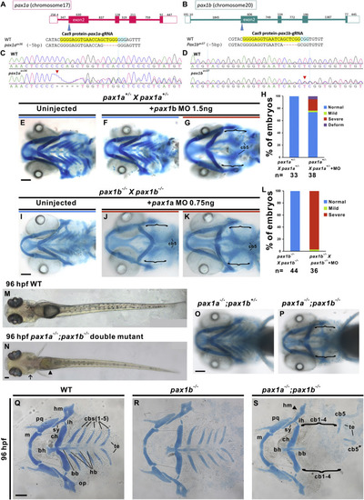
Fig. 5
pax1a- and pax1b-deficient embryos generated by CRISPR-Cas9 mutagenesis exhibit abnormality of hyoid cartilage and lack of ceratobranchial cartilages 1–4. Genomic structures of pax1a (A) and pax1b (B) include the target sites for pax1a sgRNA and pax1b sgRNA in exon 2 of each gene. Nucleotide sequence of wild type (WT) and sequences of pax1aas36 and pax1bas37 mutants containing 5-bp deletions (red dashed line) are shown (A, B). sgRNA sequence (yellow highlight) and protospacer adjacent motif (PAM, blue highlight) are indicated. (C, D) Sequences of WT and pax1aas36 or pax1bas37 mutants were compared. Positions of deleted sequences in pax1aas36 or pax1bas37 mutants are indicated by arrowheads. Normal patterning of pharyngeal arches was detected in Alcian blue-stained uninjected (E) or 1.5 ng pax1b MO-injected pax1a+/− incrossed embryos (F) at 96 hpf. About 21.1% of pax1a+/− incrossed embryos that had been injected with 1.5 ng pax1b MO failed to develop ceratobranchial cartilages (G, H). Similarly, normal pharyngeal cartilage development was identified in Alcian blue-stained uninjected pax1b−/− incrossed embryos (I) while loss of ceratobranchial cartilage and hyoid cartilage with inverted (J) or no (K) anterior-posterior polarity were detected in 0.75 ng pax1a MO-injected pax1b−/− incrossed embryos stained with Alcian blue at 96 hpf. A comparison of the percentage embryos with pharyngeal cartilage defects in uninjected and pax1a MO-injected pax1b−/− incross embryos is shown (L). A pax1a−/−; pax1b−/− double mutant embryo (N) exhibits a straight body, lack of swim bladder (arrowhead) and lack of gill cartilages (arrow), in contrast with wild type (M) at 96 hpf. Alcian blue-stained pax1a−/−; pax1b−/− double mutant embryo (P) has no ceratobranchial cartilage, in contrast with pax1a−/−; pax1b+/− mutant embryo (O) with normal pharyngeal cartilages at 96 hpf. Flat mount Alcian blue staining of pharyngeal cartilage revealed loss of ceratobranchial (cb) cartilages 1–4 with retention of shorter cb cartilage 5 in pax1a−/−; pax1b−/− double mutants. Basibranchial (bb) cartilages 2–5 were also absent, with retention of bb cartilage 1. All hypobranchial (hb) cartilages and anterior hyomandibula (hm, arrowhead) were also absent in pax1a−/−; pax1b−/− double mutants (S) in contrast to wild types (Q) and pax1b−/− mutant embryos (R) at 96 hpf. Brackets indicate lack of cb1–4. Ch, ceratohyal; ih, interhyal; m, Meckel's cartilage; op, opercular bone; pq, palatoquadrate; sy, symplectic; te, teeth. Scale bars represent 100 μm. |
| Fish: | |
|---|---|
| Knockdown Reagents: | |
| Observed In: | |
| Stage: | Day 4 |
Reprinted from Mechanisms of Development, 161, Liu, Y.H., Lin, T.C., Hwang, S.L., Zebrafish Pax1a and Pax1b are required for pharyngeal pouch morphogenesis and ceratobranchial cartilage development, 103598, Copyright (2020) with permission from Elsevier. Full text @ Mech. Dev.