|
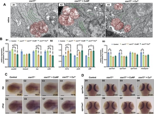
Retina development in cox17−/− mutant treated with copper. A TEM analysis of retinal cells in copper stressed cox17−/− mutant embryos at 96 hpf. A1-A3, sagittal slides of retina, red color indicating mitochondria and green color indicating ER. B Quantitative PCR of ROS related genes (B1; cox4i2, hmox2, lox, and nrf2), UPR related genes (B2; ire1a, perk, and atf6), and retinal genes (B3; opn1wl1, opn1sw1, opn1sw2 and rhodopsin) in cox17−/− mutants. C WISH assays of ER stress marker bip and chop in cox17−/− mutants. D WISH assays of retinal marker genes (opn1wl1 and opn1sw1) in cox17−/− mutants. C1-C8, lateral view, anterior to the left, D1-D8, dorsal view, anterior to the up, and Scale bar: A1-A3, 1 μm; C1-C8 and D1-D8, 100 μm. **, P < 0.01; *, P < 0.05
|