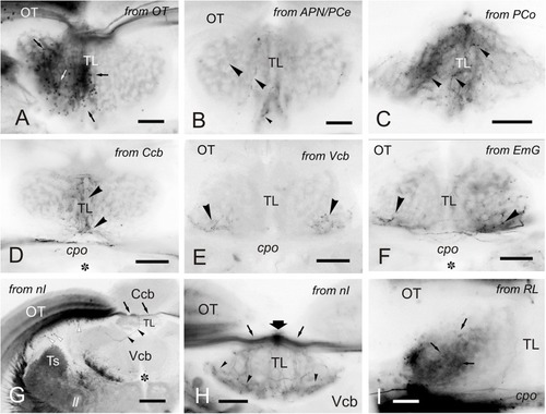
Labeled cells and fibers in TL after DiI application to various regions and nuclei. (A–I), Labeled structures in the TL after unilateral DiI application to the optic tectum (A) pretectal nuclei (B,C), cerebellum (D-F), nucleus isthmi (G,H) and nucleus rostrolateralis (I). (A) Small labeled cells in the ipsilateral TL (arrows) after tracer application to the OT. (B) Very few labeled fibers (arrowheads) in the medial TL after DiI application to the APN and PCe region. (C) Labeled fibers (arrowheads) in the medial TL after tracer application to the paracommissural nucleus. (D–F) Bilaterally labeled fibers (arrowheads) in the ventrolateral TL after unilateral tracer application to the cerebellar corpus (D), cerebellar valvula (E), and granular eminence (F). (G,H) Labeled fibers (arrowheads) after unilateral DiI application to the nucleus isthmi region. Note labeled cells in the optic tectum (white arrowheads) and labeled fibers coursing the intertectal commissure dorsal to TL (thin arrows) in (G) and (H), so as a dorsal longitudinal fascicle (thick arrow) in (H). Note also some fibers in the ipsilateral torovalvular tract (arrowheads), likely labeled by DiI diffusion to nucleus isthmi neighboring areas (see Section “DISCUSSION”), and bilateral labeled fibers in the TL (H). (I), Ipsilateral faintly labeled cells (arrows) in the TL from the rostrolateral nucleus region Asterisk: ventricle. For abbreviations, see the list. Scale bars: Scale bars: 200 μm (G); 100 μm (A-F,H); 20 μm (I).
|
