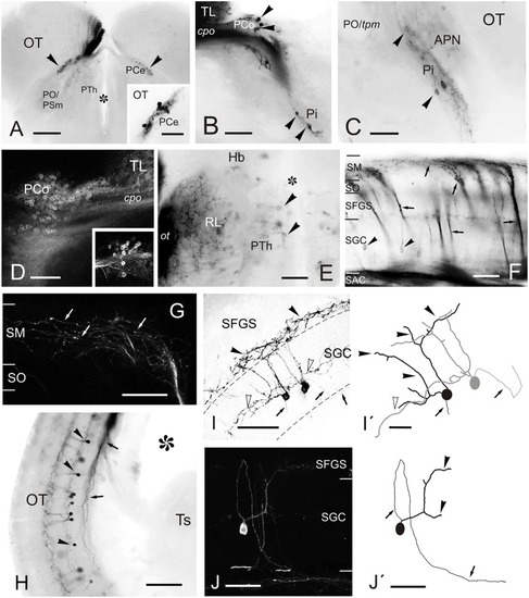
Labeled cells and fibers after direct DiI application to TL. (A–J) Photomicrographs of labeled structures observed in transverse sections at diencephalic and mesencephalic levels after DiI application to the TL. (A) Labeled cells in the central pretectal nucleus (PCe; arrowheads). Inset: detail of labeled cells in the PCe. (B,C) Retrogradely labeled cells (arrowheads) in the intercalated (Pi; in B and C) and paracommissural (PCo; in B) pretectal nuclei. (D) Confocal image showing retrogradely labeled cells in the ipsilateral PCo. Inset: contralateral PCo. (E) Faintly labeled cells (arrowheads) in the prethalamus and labeled terminals and fibers in the nucleus rostrolateralis and optic tract. (F) Retrogradely labeled tectal cells (black arrowheads) and anterogradely labeled bundles of marginal axons (arrows) through the optic tectum to reach the marginal layer (SM) where they spread giving beaded axon profiles. (G) Confocal image of the marginal layer showing a detail of beaded parallel fibers (arrows) spreading from one torofugal tract. (H) Retrogradely labeled cells (black arrowheads) and fibers (arrows) in the tectum. (I) Inverted image of the projection of a confocal stack with two fluorescent toropetal tectal cells showing dendritic arborization in the SGC (open arrowheads) and ventral SFGS (black arrowheads) and, their axon directed ventrally to the SAC (arrows). (I’) Tracing of the cells shown in (I). (J) Confocal image of a toropetal tectal cell. (J’) Tracing of cell shown in (J). Asterisk, ventricle. For abbreviations, see list. Scale bars: 200 μm (A); 100 μm (B,H); 50 μm (D,G,I–J,J’); 20 μm (inset in A,C,E,F,I’).
|
