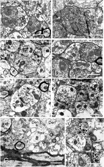
Fine cell processes structure of the adult TL. (A–H), Electron micrographs of the TL neuropil showing different axon terminals and dendrites. (A) Type I axon terminal (at1), with a number of small vesicles and large mitochondria (m) besides a dendritic process (Den) with smaller mitochondria (asterisk). Arrows: wavy-shaped synaptic active zone. (B) Type I axon terminal (at1) densely filled with vesicles in close contact with a dendrite (Den). Arrows: wavy-shaped synaptic active zone. Note that mitochondria in the post-synaptic dendrite (asterisk) are smaller and more abundant. (C) Axon terminal type I (at1) making synapse (arrows) with two dendritic processes (Den1, Den2). (D) Axo-axonic contact between type I (at1) and type II (at2) terminals, as well as a type I axo-dendritic synapse (arrows). (E) An axo-dendritic synapse (arrow) for a type II axon terminal. Note a myelinated axons (axm) on the top-right and left. (F) Axo-axonic synapse (arrows) between type I (at1) and II (at2) terminals, as well as axo-dendritic synapses (arrows) with two dendritic processes (Den1, Den2). (G) Axon terminal type III (at3) in the rostral TL intermingled with myelinated (axm) and unmyelinated (ax) axons and dendrites (Den). Note the thick and highly myelinated axons in the posterior commissure at the bottom of the image (star). (H) Detail of axon terminals type III (at3), with small clear (black arrows) and large electron-dense vesicles (white arrows), next to type I (at1) axon terminals. m, mitochondria. Scale bars: 1 μm (A–F,H); 2 μm (G).
|
