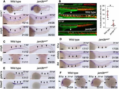
Mutation of jam3b causes defects in both hematopoietic and vascular development. (A) Expression of runx1 and cmyb in the DA of WT or jam3bsa37 embryos. Magnification of boxed area shown underneath. (B) Representative image (left) and quantification (right) of cd41:GFP; kdrl:mCherry double-positive cells in WT or jam3bsa37 embryos. White arrowheads indicate double-positive cells in the ventral floor of the DA. Magnification of boxed area shown underneath. Data are mean±s.d. *P<0.00001 (unpaired two-tailed Student's t-test). (C) Expression of cmyb in the CHT and rag1 in the thymus of WT or jam3bsa37 embryos. (D) Expression of fli1a (endothelium), efnb2a (DA), and flt4 (PCV) in WT or jam3bsa37 embryos. (E) Expression of gata1a and hbae3 in primitive erythrocytes of WT or jam3bsa37 embryos. (F) Expression of fli1a and l-plastin (macrophage) in WT or jam3bsa37 embryos. Black arrowheads indicate the expression domain of each gene. Numbers in bottom right of panels indicate the number of embryos showing the displayed expression pattern over the total number of analyzed embryos. Scale bars: 100 μm.
|
