
Figure 2—figure supplement 3.
- ID
- ZDB-FIG-200220-40
- Publication
- He et al., 2020 - In vivo single-cell lineage tracing in zebrafish using high-resolution infrared laser-mediated gene induction microscopy
- Other Figures
- All Figure Page
- Back to All Figure Page
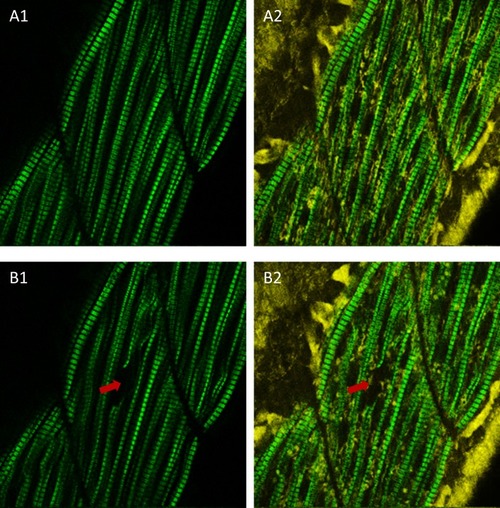
Laser-induced injury in zebrafish muscle.(A1) SHG image of zebrafish muscle before laser heating. (A2) Merged image of SHG and reduced nicotinamide-adenine dinucleotide (NADH) autofluorescence signals of zebrafish muscle before laser heating. (B1) SHG image of zebrafish muscle after focusing 85 mW IR laser to a single fiber for heating over 3 s. (B2) Merged image of SHG and NADH autofluorescence signals of zebrafish muscle after IR laser heating. The arrows in (B) indicate the laser heating point, where the sarcomere structure disappeared, indicating that muscle fiber was injured. To optimize the heat shock gene induction and avoid cell damage, scan heating should be performed for in vivo heat shock in zebrafish. The field of view of (A) and (B): 100 µm × 100 µm. |