|
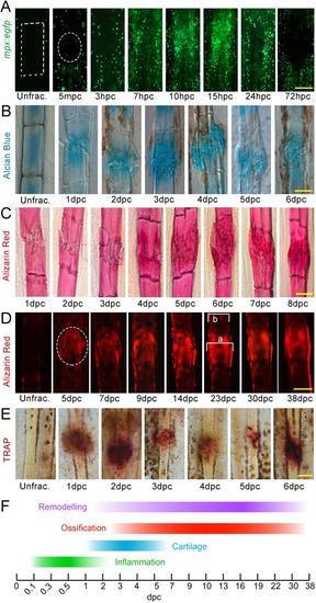
Phases of bone crush fracture repair in the adult zebrafish fin. (A-E) Fluorescent (A,D) and brightfield (B,C,E) images of mpx:egfp (A), Alcian Blue (B), Alizarin Red (C,D) and TRAP (E) labelling of lepidotrichia crush fractures at different stages of repair. Unfractured stainings are shown for reference. GFP-positive neutrophils (A), soft callus (B), hard callus (C,D) and osteoclast remodelling (E) have distinct temporal properties during repair. Fluorescent imaging of the bone callus (D) demonstrates the presence of a bone collar surrounding the callus. Relative callus width was measured by dividing the width of callus by the width of adjacent lepidotrichia (a/b) (D). Crush positions in A and D are circled; ray segment is outlined by a box in A. (F) Scheme showing the stages of repair. Scale bars: 100 µm. mpc, minutes post crush.
|