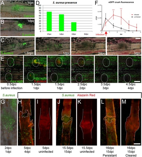
S. aureus infection of fractures delays healing. (A-C) Fluorescent images overlying Nomarski images of either calcein tracer (A) or eGFP-labelled S. aureus (B,C), showing that both can be successfully introduced to a fracture site. (D) Injection of 0.5 nl of 2500 cfu/nl S. aureus yielded detectable bacteria up to 3 dpi (n=20 per point). (E) Fluorescent images of mpx:egfp-positive neutrophils (green) and mCherry-expressing S. aureus (red) over 4 days of infection introduced 12 h after crushing. (F) Inflammation correlates with the presence of S. aureus and prolongs inflammatory response at the fracture (n=4 per point). ***P<0.001; ANOVA with Sidak post test. (G-M) Fluorescent images of eGFP-expressing S. aureus introduced either 12 (J) or 24 h (G,H,L,M) following fracture and viewed with either Nomarski optics (G) or with fluorescence for Alizarin Red (H-M). (I,K) Corresponding uninfected fracture controls are shown. Outlines indicate bone stained by Alizarin Red, showing that the presence of S. aureus predicts poor callus formation compared with uninfected controls (I,K) or where the infection has been cleared (M). Scale bars: in C and G, 100 µm for A-C,G-M; E, 200 µm.
|
