IMAGE

Fig. 1.

ID
ZDB-IMAGE-191230-633
Source
Figures for Tomecka et al., 2019
Image
Figure Caption

Fig. 1.

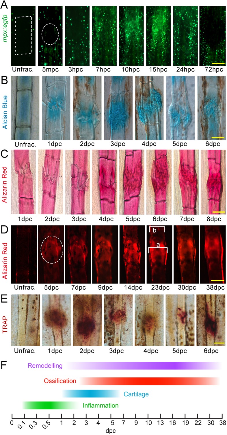
Phases of bone crush fracture repair in the adult zebrafish fin. (A-E) Fluorescent (A,D) and brightfield (B,C,E) images of mpx:egfp (A), Alcian Blue (B), Alizarin Red (C,D) and TRAP (E) labelling of lepidotrichia crush fractures at different stages of repair. Unfractured stainings are shown for reference. GFP-positive neutrophils (A), soft callus (B), hard callus (C,D) and osteoclast remodelling (E) have distinct temporal properties during repair. Fluorescent imaging of the bone callus (D) demonstrates the presence of a bone collar surrounding the callus. Relative callus width was measured by dividing the width of callus by the width of adjacent lepidotrichia (a/b) (D). Crush positions in A and D are circled; ray segment is outlined by a box in A. (F) Scheme showing the stages of repair. Scale bars: 100 µm. mpc, minutes post crush.

Acknowledgments
This image is the copyrighted work of the attributed author or publisher, and ZFIN has permission only to display this image to its users. Additional permissions should be obtained from the applicable author or publisher of the image. Full text @ Dis. Model. Mech.