
Figure 3
- ID
- ZDB-FIG-191230-1816
- Publication
- Tang et al., 2019 - Cardiac neural crest contributes to cardiomyocytes in amniotes and heart regeneration in zebrafish
- Other Figures
- All Figure Page
- Back to All Figure Page
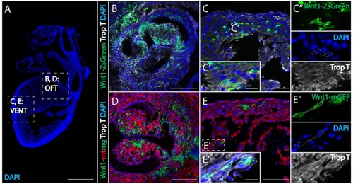
(A) Low magnification image to show the relative anatomical positions of a mouse heart at E15.5 (sagittal view, DAPI-blue). (B, C) In Wnt1-Cre; ZsGreenfl/fl mice, neural crest-derived cells (green, Wnt1-Cre driven ZsGreen expression is abbreviated as Wnt1-ZsGreen, enclosed in dashed line) were observed in myocardium (Troponin T, gray) of the outflow tract (B), and ventricle (VENT) (C, C’’: separate channels of inset C’). (D, E) Similar results were obtained from Wnt1-Cre2+; R26mTmG mice (Wnt1-Cre2+ driven replacement of membrane localized tdTomato (mT) by EGFP (mG) (abbreviated as Wnt1-mtmg), where cardiac crest-derived cells (green, enclosed in dashed line) were present in myocardium of the outflow tract (D) and ventricle (Troponin T, gray) (E, E’’: separate channels of inset E’). Transverse view. Spatial-temporal information and antibody staining are displayed in supplement 1. Scale bars: A 400 μm; B-E 100 μm; C’, E’ 10 μm. |