
Figure 2
- ID
- ZDB-FIG-191230-1665
- Publication
- Fillatre et al., 2019 - TEADs, Yap, Taz, Vgll4s transcription factors control the establishment of Left-Right asymmetry in Zebrafish
- Other Figures
- All Figure Page
- Back to All Figure Page
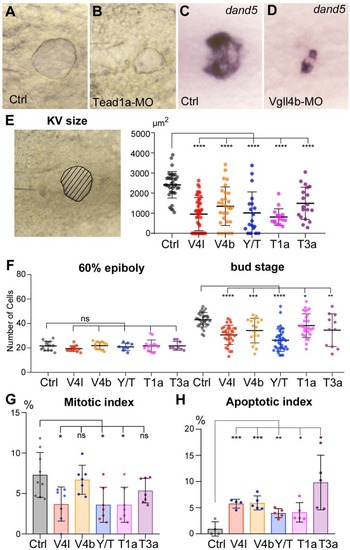
( A–D) Illustration of the strong decrease in the size of the KV at the 12-somite stage in loss-of-function conditions shown in brightfield for ( A) a control embryo (Ctrl) and for ( B) a TEAD1a morphant embryo (Tead1a-MO) and by in situ hybridization using a dand5 probe in ( C) Ctrl and in ( D) Vgll4b morphant embryo (Vgll4b-MO). ( E–H) Effect of Vgll4l (V4l), Vgll4b (V4b), Tead1a (T1a), Tead3a (T3a) loss of function and of Yap/Taz (Y/T) double loss of function on: ( E) the size of the KV (expressed as the area of the planar projection of its lumen), ( F) the number of DFCs present at early gastrula stage (60% epiboly) and at the end of gastrulation (bud stage), ( G) the proliferation of the DFCs measured as their mitotic index at 75% of epiboly, ( H) the survival of DFCs measured as their apoptotic index at 90% epiboly. In all cases control (Ctrl) embryos were injected with 8 ng of Standard MO. Graph indicates the mean of each experiment, error bars indicate standard deviation and dots indicate the individual measurement for DFC groups or individual KV in control and loss of function conditions. Statistical significance between controls and the different loss-of-function conditions: two-tailed unpaired t-test. *p≤0.05, **p≤0.01, ***p≤0.001, ****p≤0.0001. ns: not significant. Numerical data for ( E–H) and details of statistical analysis are provided in Figure 2—source data 1. |
| Gene: | |
|---|---|
| Fish: | |
| Knockdown Reagents: | |
| Anatomical Term: | |
| Stage: | 10-13 somites |
| Fish: | |
|---|---|
| Knockdown Reagents: | |
| Observed In: | |
| Stage Range: | 75%-epiboly to 10-13 somites |