|
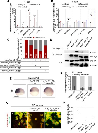
Up-regulation of the MARCKS family members and its interaction protein Hsp70.3 over-compensates the genetic loss of <italic>marcksb</italic>.(A, B) RT-qPCR analysis on the expression of marcksa, marcksb, marcksl1a, marcksl1b and hsp70.3 in the embryos indicated in the Fig at 1-cell stage (A) and shield stage (B). The data were presented as scatter plots with bar representing median value relative to respective transcript levels measured in wildtype embryos. “*”: P < 0.01, from Student’s t-test. (C) Overexpression of marcksa, marcksl1a, marcksl1b or hsp70.3 partially rescued the dorsalization defects in marcksb morphants. “n” represents the number of embryos we observed. (D) Co-immunoprecipitation revealed that Hsp70.3 had high binding affinity with Marcksb and moderate binding affinity with Marcksl11a and Marcksl1b, but relative low binding affinity with Marcksa. TCL: total cell lysis. The molecular mass of Marcks-myc is around 70KD. (E) The expression of szl was remarkably reduced in MZmarcksb injected with either hsp70_MO or marcksa_l1a_l1b_MO. The embryos are at shield stage and lateral view with dorsal to the right. (F) The percentage of embryos with normal-like and severely decreased expression of szl. “n” represents the number of embryos we observed. (G) Hsp70 interacts with Marcksa, Marcksl1a and Marcksl1b to maintain sufficient level of extracellular Bmp2b in MZmarcksb. (a-c) Compared with MZmarcksb embryos (a), knockdown of either hsp70 (b) or a combination of marcksa, marcksl1a and marcksl1b (c) remarkably reduced the level of extracellular Bmp2b in MZmarcksb. (H) Quantitative measurement of secreted Bmp2b in embryos of MZmarcksb, MZmarcksb injected with either hsp70_MO or marcksa_l1a_l1b_MO. The data were presented as scatter plots with median; “*”: P < 0.01, from Student’s t-test.
|